Biochimica et Biophysica Acta (BBA) – General Subjects, 2019, pp. 129485
DOI:10.1016/j.bbagen.2019.129485
Abstract
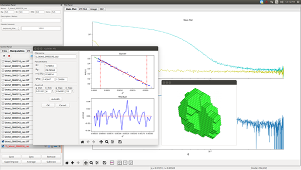
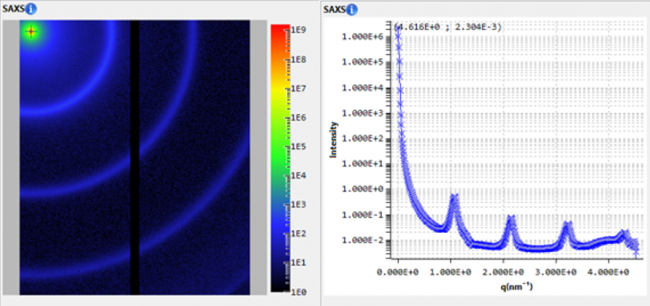
Microgels offer opportunities for improved delivery of antimicrobial peptides (AMP). To contribute to a foundation for rational design of such systems, we here study the effects of electrostatics on the generation of peptide-carrying microgels. For this, alginate microgels loaded with polymyxin B and cross-linked by Ca2+, were formed by electrostatic complexation using a hydrodynamic focusing three-dimensional (3D)-printed micromixer, varying pH and component concentrations. The structure of the resulting composite nanoparticles was investigated by small-angle X-ray scattering, dynamic light scattering, and z-potential measurements, whereas peptide encapsulation and release was monitored spectrophotometrically. Furthermore, membrane interactions of these systems were assessed by dye leakage assays in model lipid vesicles. Our results indicate that charge contrast between polymyxin B and alginate during microgel formation affects particle size and network dimensions. In particular, while microgels prepared at maximum polymyxin B-alginate charge contrast at pH 5 and 7.4 are characterized by sharp interfaces, those formed at pH 9 are characterized by a more diffuse core, likely caused by a weaker peptide-polymer affinity, and a shell dominated by alginate that shrinks at high CaCl2 concentrations. Quantitatively, however, these effects were relatively minor, as were differences in peptide encapsulation efficiency and electrolyte-induced peptide release. This demonstrates that rather wide charge contrasts allow efficient complexation and particle formation, with polymyxin B encapsulated within the particle interior at low ionic strength, but released at high electrolyte concentration. As a consequence of this, peptide-mediated membrane destabilization were suppressed by microgel incorporation at low ionic strength, but regained after microgel disruption. After particle disruption at high ionic strength, however, some polymyxin B was found to remain bound to alginate chains from the disrupted composite microgel particles, resulting in partial loss in membrane interactions, compared to the free peptide.