Soft Matter, 2019, vol 15, 23, pp. 4751-4760
DOI:10.1039/C9SM00615J
Abstract
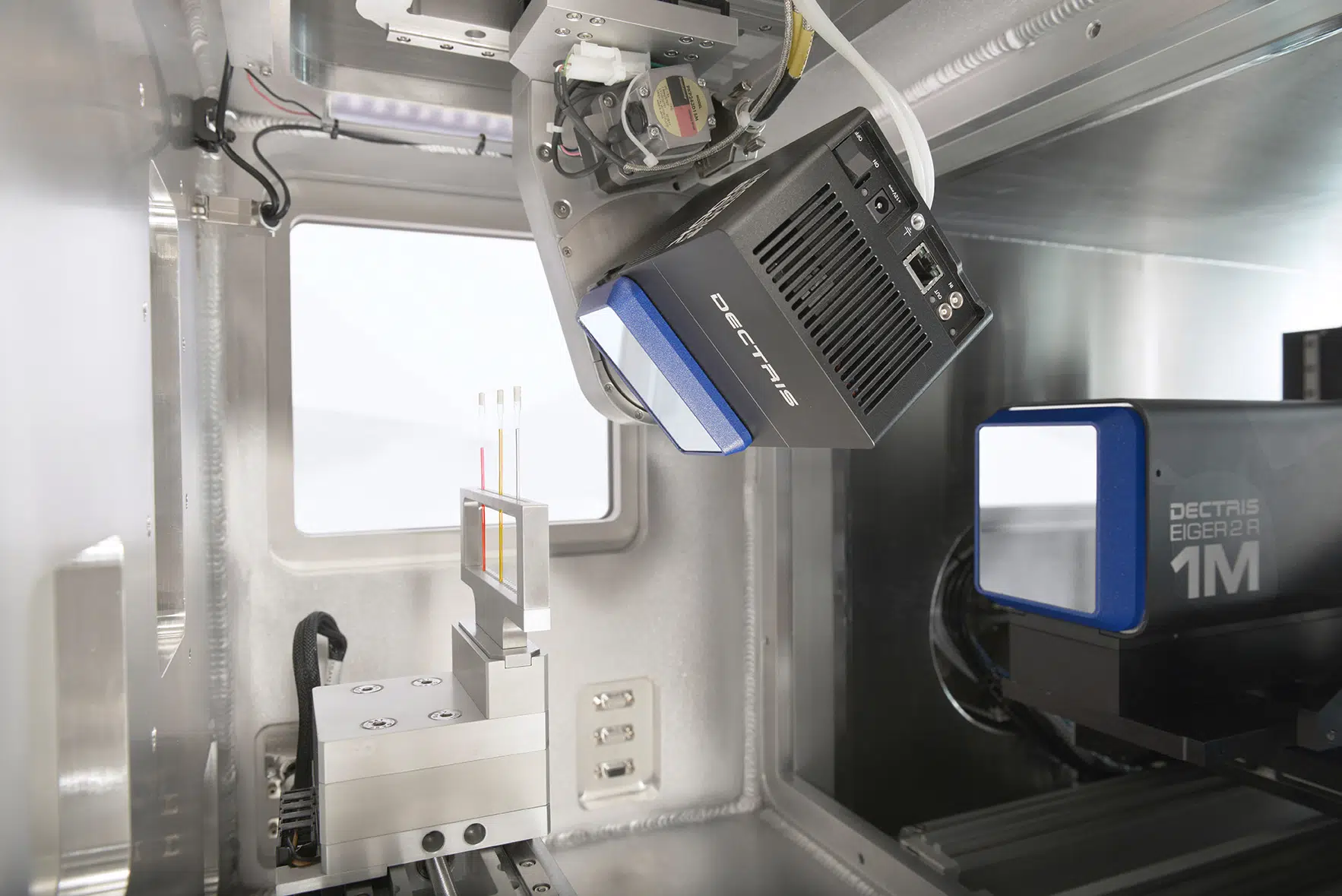
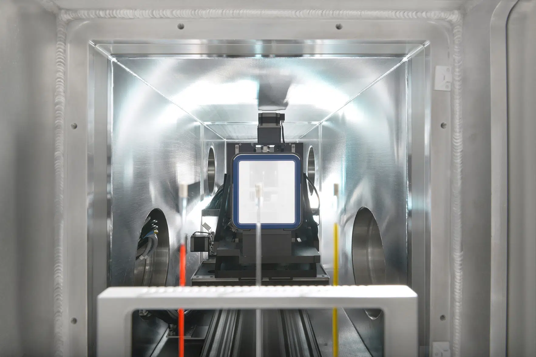
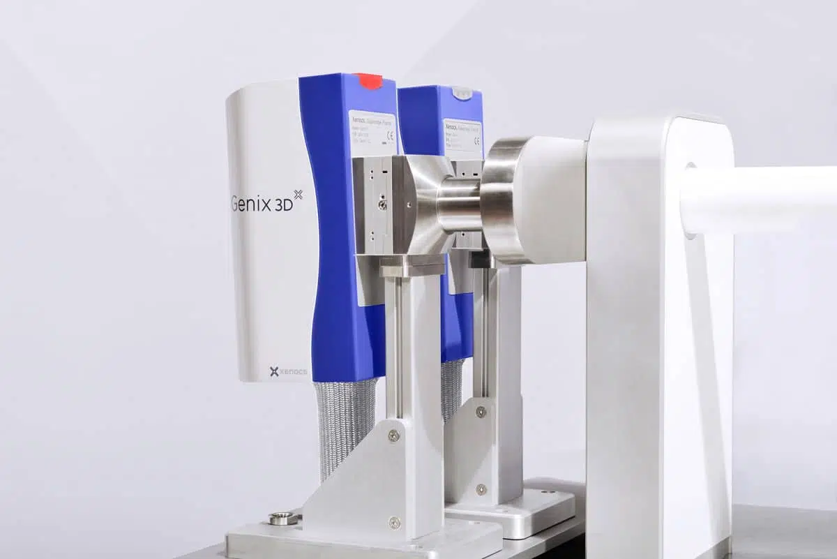
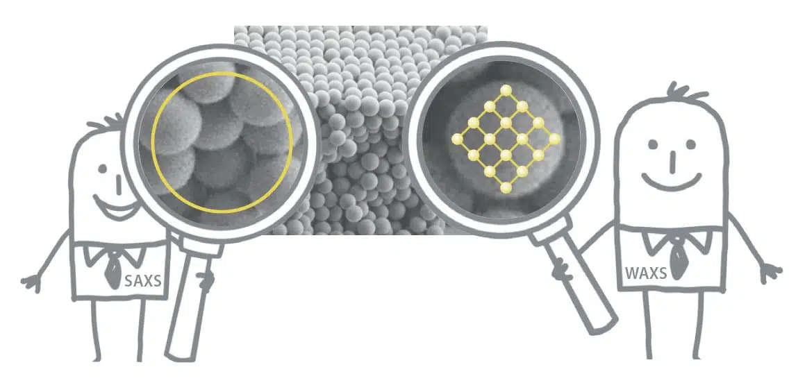
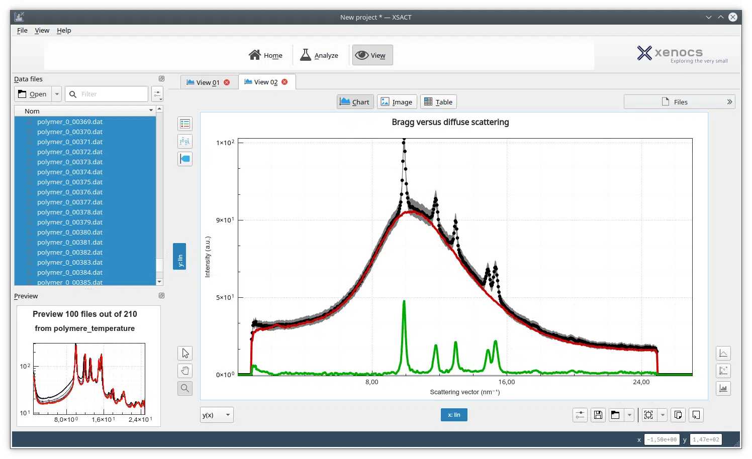
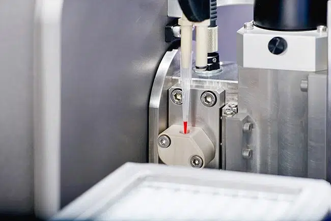
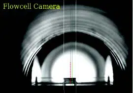
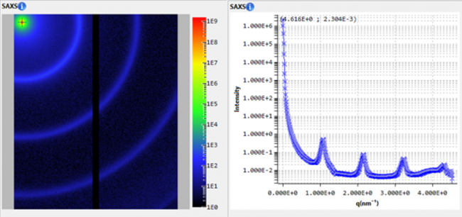
It has been previously reported that poly(ethylene) (PE)-based block copolymers self-assemble in certain thermosetting matrices to form a dispersion of one-dimensional (1D) nanoribbons. Such materials exhibit exceptional properties that originate from the high aspect ratio of the elongated nano-objects. However, the ability to prepare 1D assemblies with well-controlled dimensions is limited and represents a key challenge. Here, we demonstrate that the length of ribbon-like nanostructures can be precisely controlled by regulating the mobility of the matrix during crystallization of the core-forming PE block. The selected system to prove this concept was a poly(ethylene-block-ethylene oxide) (PE-b-PEO) block copolymer in an epoxy monomer based on diglycidyl ether of bisphenol A (DGEBA). The system was activated with a dual thermal- and photo-curing system, which allowed us to initiate the epoxy polymerization at 120 °C until a certain degree of conversion, stop the reaction by cooling to induce crystallization and micellar elongation, and then continue the polymerization at room temperature by visible-light irradiation. In this way, crystallization of PE blocks took place in a matrix whose mobility was regulated by the degree of conversion reached at 120 °C. The mechanism of micellar elongation was conceptualized as a diffusion-limited colloid aggregation process which was induced by crystallization of PE cores. This assertion was supported by the evidence obtained from in situ small-angle X-ray scattering (SAXS), in combination with differential scanning calorimetry (DSC) and transmission electron microscopy (TEM).