What are the advantages & drawbacks of small angle X-ray scattering (SAXS) and transmission electron microscopy (TEM), and how do they complement each other?
The field of nanotechnology has seen a steady increase in interest from both public research institutions and commercial companies over the last two decades [1, 2]. Nowadays nanomaterials find applications in a plethora of sectors from biomedicine to microelectronics, food science and agriculture. These materials exhibit different electronic, optical, mechanical and chemical properties from their bulk counterparts [3]. Thus, comprehensive, accurate and reliable nanoscale level characterization plays a crucial role in the understanding and development of these materials. However, due to the inherent difficulties encountered in studying nanomaterials, it is oftentimes necessary to combine two or more characterization methods to obtain a full picture of the variety of properties associated with a particular system. Therefore, in order to choose and combine effectively the right methods, it is important to understand the strengths and limitations of the different techniques. Here we focus on presenting the roles of small angle X-ray scattering (SAXS) and transmission electron microscopy (TEM) in a comparative way with the aim to understand their unique capabilities as well as their complementarities.
To begin with, the two characterization methods are based on fundamentally different working principles. SAXS is an indirect method that makes use of X-rays to generate a scattering pattern that is interpreted in the reciprocal space. In contrast, TEM is a direct method that produces an image in real space by means of electron beam imaging.
SAXS
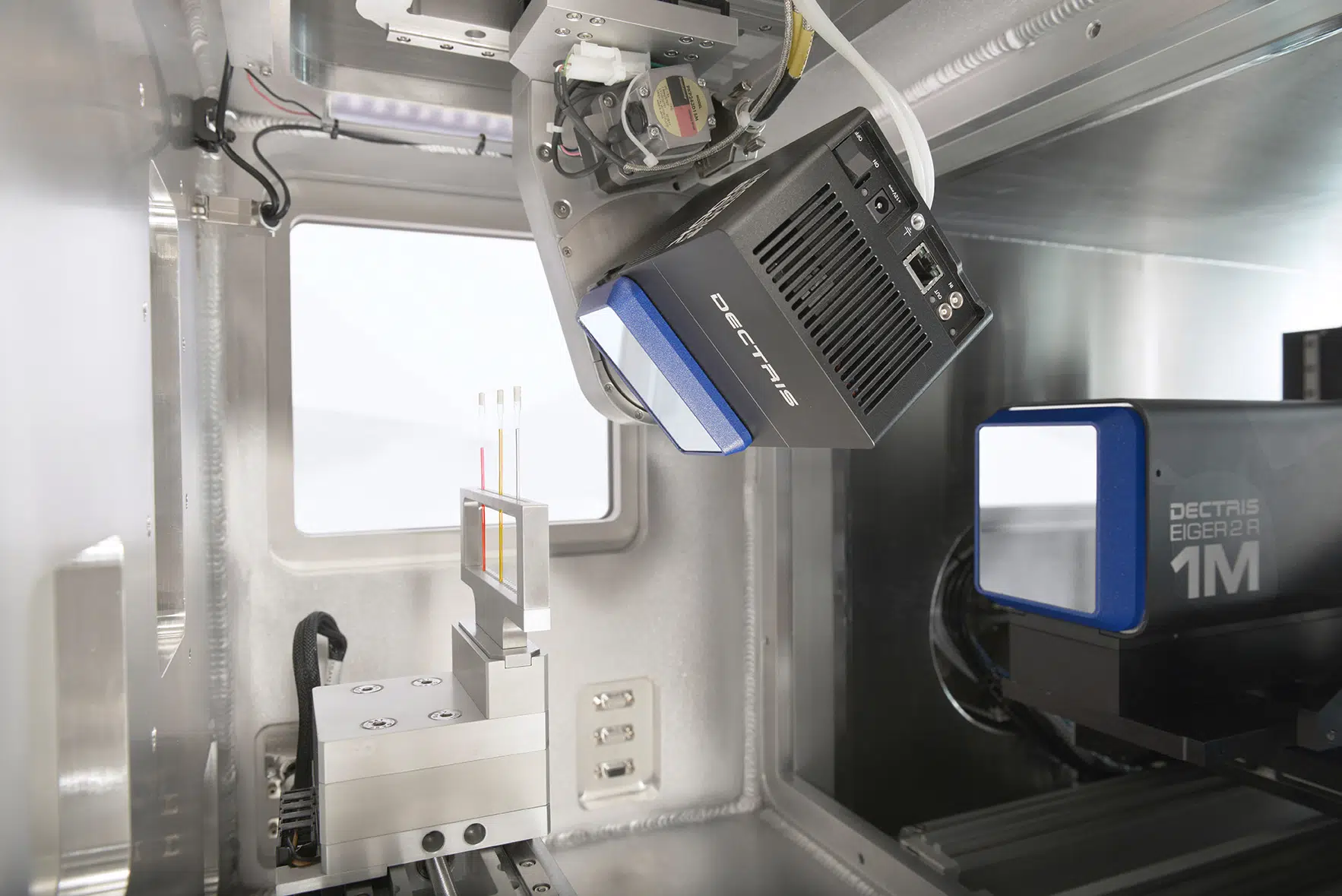
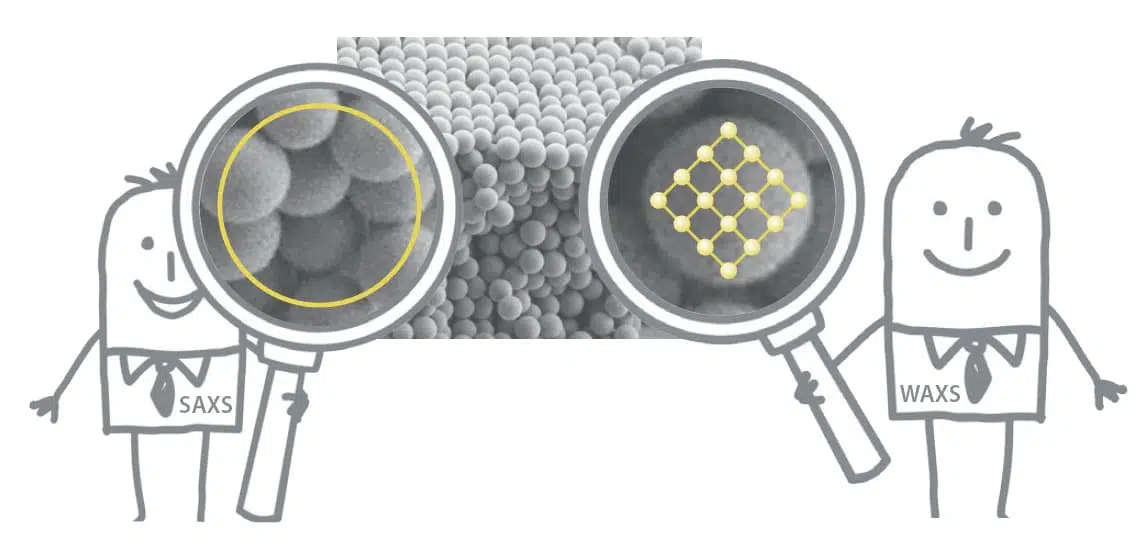
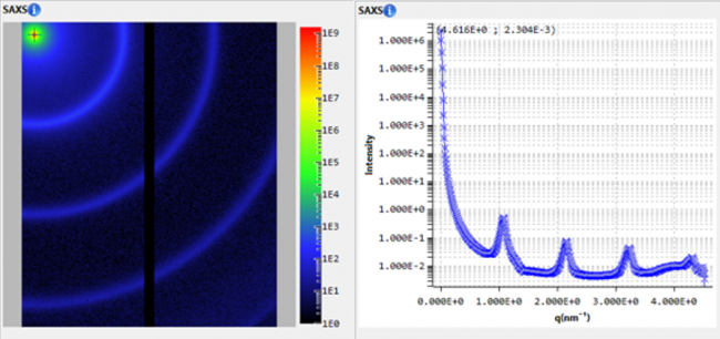
As the name suggests, during a SAXS measurement the sample is illuminated by a monochromatic beam of X-rays and scattered photons are recorded down to low angles (typically below 10o). The angular dependence of the intensity contains a multitude of information such as the size and size distribution of particles and pores, particle and macromolecular shape or crystalline and surface structure. By making use of the different available angular ranges (wide, small, and ultra-small angles WAXS/SAXS/USAXS) and geometries (transmission mode or grazing incidence GISAXS) one can extract sub-nanometer up to micrometer structural information both from bulk samples and their surfaces.
One particular advantage of SAXS is its capability to probe a large volume with respect to the particle size, providing statistically relevant results, e.g., a typical observation volume of 1 mm3 would contain several billions of 100 nm particles, and all orientations of the particles will be probed. The drawback of this method is a reduced spatial resolution and lack of local information. Additionally, there are fewer restrictions imposed by the use of energetic x-ray (typically in the range of 8 keV) thus offering the possibility to probe samples in various forms (liquid, solid, powder, etc.) with limited sample preparation requirements. Compared to imaging techniques, for instance TEM, the data analysis for a SAXS experiment is more complex and generally requires some minimal prior knowledge about the sample morphology, such as chemical or shape hypotheses, to avoid misinterpretations.
TEM
TEM makes use of a high voltage to accelerate electrons which are transmitted through a thin sample and collected to create a 2D projection of the materials’ nanoscale structure. Being performed in transmission mode additional restrictions are imposed on the sample. Besides the request for ultra-thin sections (typically 100 nm thick) samples also need to be uniform, electron transparent, free from contamination (especially carbon contamination [4]), and in a form suitable for high vacuum operation. Ultimately this results in a complicated and time-consuming sample preparation step, often far from real sample environment in bulk, solutions etc.
However, once the right sample is in the beam, TEM is able to produce some of the highest resolution structural analysis and eventually also provide elemental information. One of its strengths is that it offers a straightforward characterization of the shape and estimate of the particle size as well as a relative estimation of the samples’ degree of homogeneity/size dispersion. Accurate determination of particle parameters, such as size distribution, is complicated by a number of operator bias artifacts: as TEM is a local probe the choice of the analyzed section of the sample can be nonrepresentative of the entire material, the experimenter-guided inspection of single particles is time-consuming, limited to a few hundreds of particles and could favor the selection of larger particles which in general provide higher contrast, and finally, inhomogeneities can be formed during sample preparation (especially while removing the suspension liquid after dropcasting) [6]. Specific orientation of particle can be also induced at this preparation step generating wrong interpretation scenarios. Indeed, the image being formed by the projection of the object either by density contrast or phase contrast, the contribution of the 3D shape to the overall size might not be reachable except in the case of very anisotropic objects (such as discs) where an obvious orientation is observed (side of disc). Furthermore, imaging artifacts can be misleading, e.g., a population of discs can be falsely interpreted as bi population of discs and rods when it can be simply composed of a single population of discs with some of them being observed on their edge.
Therefore, depending on the goal of your research and its context, you might find that one of the techniques is sufficient to answer your questions. However, as SAXS and TEM offer complementary information, an exhaustive description of the sample properties, such as the size and shape of nanoparticles and their spatial distribution, or degree of homogeneity, can be obtained by combining the two methods. The modelization of SAXS can be done by an appropriate initialization using real space information of TEM and thus efficiently obtaining statistical data interpretation of the sample while maintaining it in natural conditions.
Particle size distribution of bimodal silica nanoparticles as revealed by SAXS and TEM
The applicability of nanoparticles in general and silica nanoparticles, in particular, is governed by several physicochemical properties such as particle size distribution, shape or stability. Both SAXS and TEM are excellent nanoparticle characterization methods that can provide accurate descriptions of monodisperse particle distributions.
However, when, it comes to bimodal or heterogeneous distributions TEM’s accuracy is deteriorated by the fact that the number of observed particles is limited to a few hundred or thousands at most. Fig. 1 shows a TEM image recorded on a sample composed of bimodal silica nanoparticles and the associated particle size distribution histograms measured on ~150 particles with a 5 nm bin size [5]. In the image, an apparent aggregation of the nanoparticles is visible.
SAXS on the other hand is very sensitive to the formation of aggregates with a clear signature on the scattering intensity visible at low values of the scattering vector q. Since no such aggregation was noticeable from the SAXS measurements, performed on the same particle system but in the native suspension form, it can be concluded that its origin lies in the sample preparation. More specifically, it occurs during the drying process while samples are deposited as a liquid drop onto the TEM grids. It is thus clear that depending on which section of the grid is used in the analysis there is a risk that the results would not be representative of the entire system and would also lead to misinterpretation of the aggregate formation.
SAXS on the other hand is able to provide a statistically relevant description of the bimodal silica nanoparticles system. As exhibited in Fig. 1 the size and ratio of the two populations are well modeled by the SAXS data. One drawback of the method is the fact that it requires a software analysis and currently available most popular data interpretation models are based on spherical particles (or particles with only one varying dimension). However, this field is rapidly evolving and new models of polydisperse analysis of anisotropic particles should be available in a near future. In the meantime, this assumption can be straightforwardly confirmed by TEM measurements, the two techniques thus complementing each other to obtain an accurate description of the system.
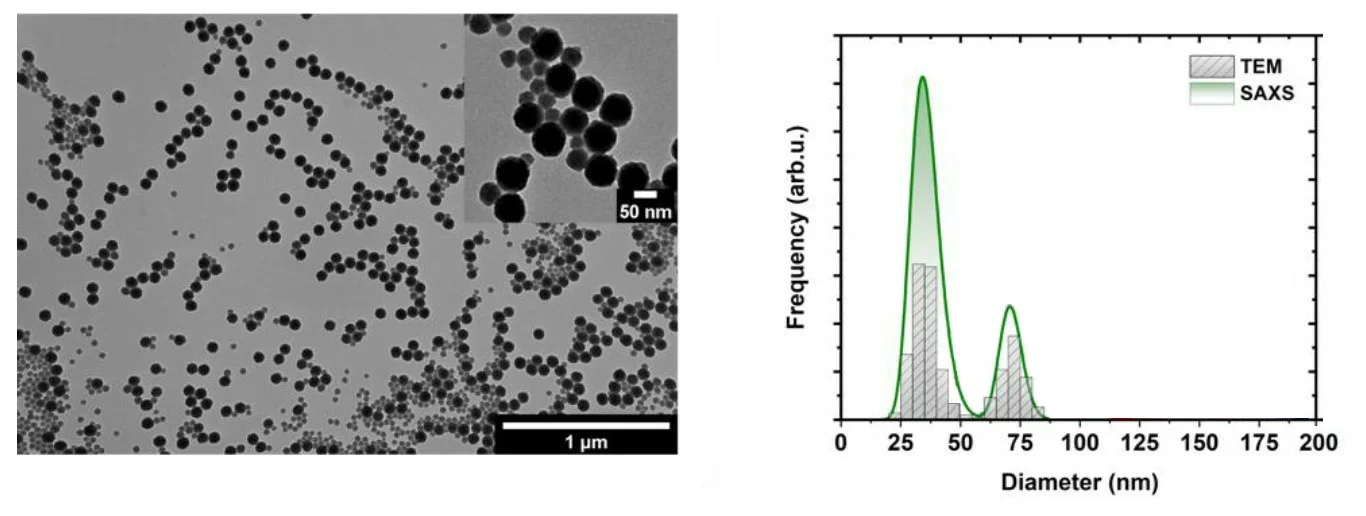
Fig. 1 Morphological characterization and particle size distributions of bimodal silicananoparticles. (left) Transmission electron microscopic (TEM) image. (right) Particle size distributions as determined by TEM and SAXS. Credit: Materials, 2020, DOI: 10.3390/ma13143101.
In conclusion, small angle x-ray scattering is a powerful routine characterization method for nanomaterials that benefits greatly from the use of microscopy techniques such as transmission electron microscopy to confirm the validity of the models used in the fitting routines.
| SAXS/WAXS GISAXS | TEM | |
| Types of samples | gel, solid, powder, liquid, turbid | Thin sections (< 100 nm) |
| Sample preparation | Minimal | Intensive |
| Volume probed | 0.1 to 1 mm^3 | Very small, thin |
| Range of size | < 1 - 350 nm (up to 2.5 µm [PP1] USAXS) | < 1 nm – 1000 nm |
| In-situ or operando | Yes | Yes |
| Measuring time | Fast | Slow |
| Statistics | Statistically relevant results | Poor |
| Spatial Resolution | Low | High |
| Data | Scattering pattern in reciprocal space | Image in real space |
| Beam damage | No | Yes |
| Limitations | Requires sufficient electron density contrast | Requires electrical conduction and high vacuum operation |
| Information | Size, shape, crystallinity, orientation, bulk nanostructures and surface analysis capability in grazing mode | Morphology based on electron density and elemental chemical contrast and possibility to use diffraction contrast |
[1] Miyazaki, Kumiko, and Nazrul Islam. “Nanotechnology systems of innovation—An analysis of industry and academia research activities.” Technovation 27.11 (2007): 661-675.
[2] Woolley, Jennifer L., and Nydia MacGregor. “Science, technology, and innovation policy timing and nanotechnology entrepreneurship and innovation.” Plos one 17.3 (2022): e0264856.
[3] Mourdikoudis, Stefanos, Roger M. Pallares, and Nguyen TK Thanh. “Characterization techniques for nanoparticles: comparison and complementarity upon studying nanoparticle properties.” Nanoscale 10.27 (2018): 12871-12934.
[4] Hettler, Simon, Manuel Dries, Peter Hermann, Martin Obermair, Dagmar Gerthsen, and Marek Malac. “Carbon contamination in scanning transmission electron microscopy and its impact on phase-plate applications.” Micron 96 (2017): 38-47.
[5] Gleichmann, Nicole. “SEM vs TEM.” Technology Networks: Analysis & Separations, online (2020): https://www. technologynetwo rks. com/analysis/articles/sem-vs-tem-331262.
[6] Yang, Ye, Suiyang Liao, Zhi Luo, Runzhang Qi, Niamh Mac Fhionnlaoich, Francesco Stellacci, and Stefan Guldin. “Comparative characterisation of non-monodisperse gold nanoparticle populations by X-ray scattering and electron microscopy.” Nanoscale 12.22 (2020): 12007-12013.
[7] Al-Khafaji, Mohammed A., Anikó Gaál, András Wacha, Attila Bóta, and Zoltán Varga. “Particle size distribution of bimodal silica nanoparticles: A comparison of different measurement techniques.” Materials 13.14 (2020): 3101.
Products
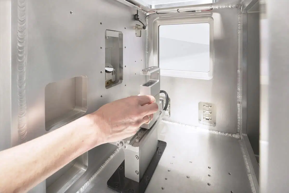
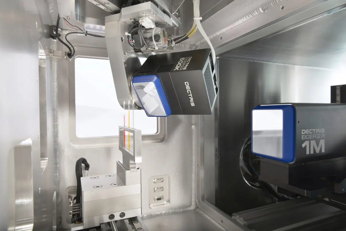
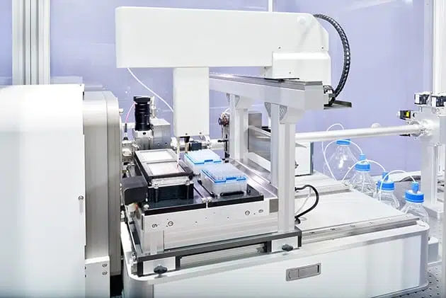
Discover our SAXS instruments in the lab
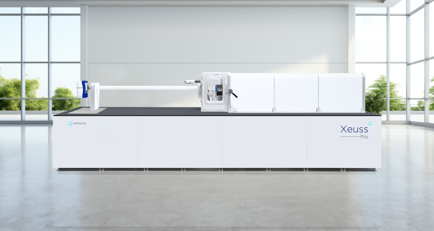
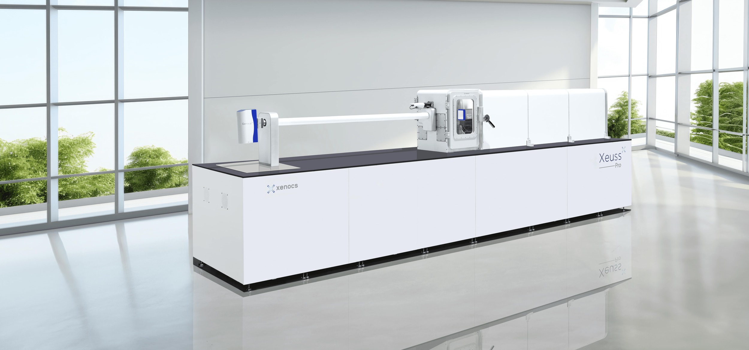
Xeuss Pro
The Ultimate Solution for Nanoscale Characterization using SAXS/WAXS/GISAXS/USAXS/Imaging
