A combined imaging and scattering approach to explore the physical origins of the dark-field signal
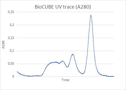
X-ray dark-field (DF) imaging provides spatial information about the presence and distribution of nano- and micrometric structural heterogeneities within a sample, such as nanoporosity, microcracks, or interfaces that remain unresolved in conventional imaging. Unlike conventional X-ray imaging, which relies on absorption contrast and is limited by detector resolution, DF imaging reveals structures smaller than the pixel size. It achieves this by detecting local variations in X-ray scattering: when X-rays interact with unresolved structures, they undergo small-angle deflections. The intensity and spatial distribution of these scattering events can then be mapped to produce an image known as a dark-field image.
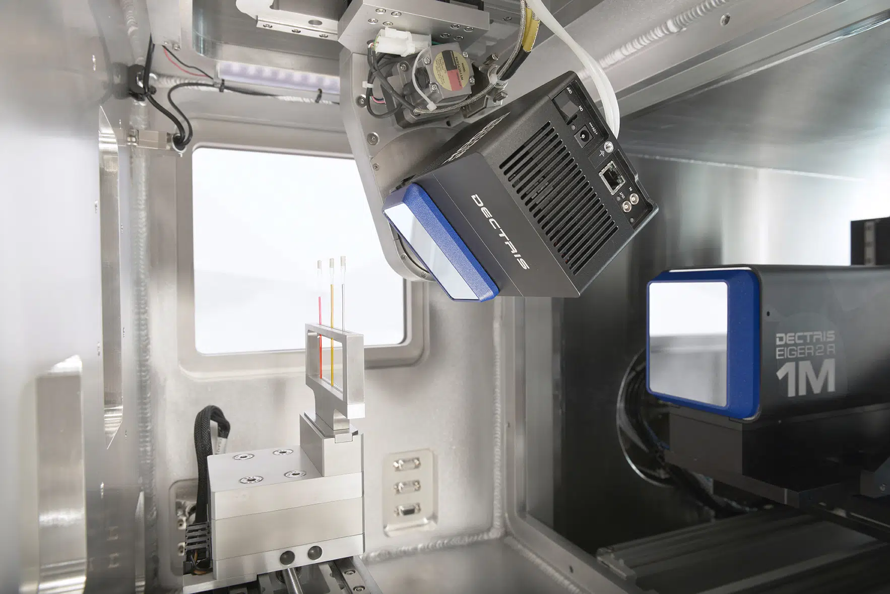
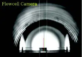
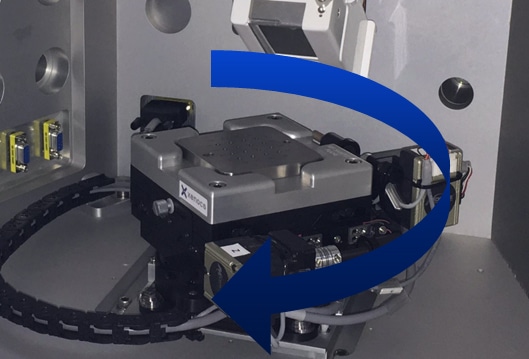
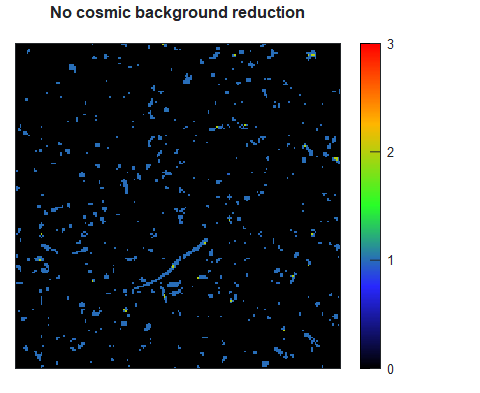
Several experimental methods and device configurations exist to produce dark-field (DF) X-ray images. These techniques, originally developed at synchrotron facilities, are based on modulating the X-ray beam upstream of the sample using optical elements such as structured masks or gratings. A reference image is first recorded using only the modulated beam; then, with the sample in place, a second image is captured. The sample induces a local blurring or loss of visibility in the modulation compared to the reference pattern. This blurring effect is then quantified numerically, enabling the reconstruction of dark-field images (see Figure 1). In laboratory setups, this principle can be implemented through the Modulation-Based Imaging (MoBI) method [1], which forms the basis of the DF-PCI option available on the Xeuss Pro.
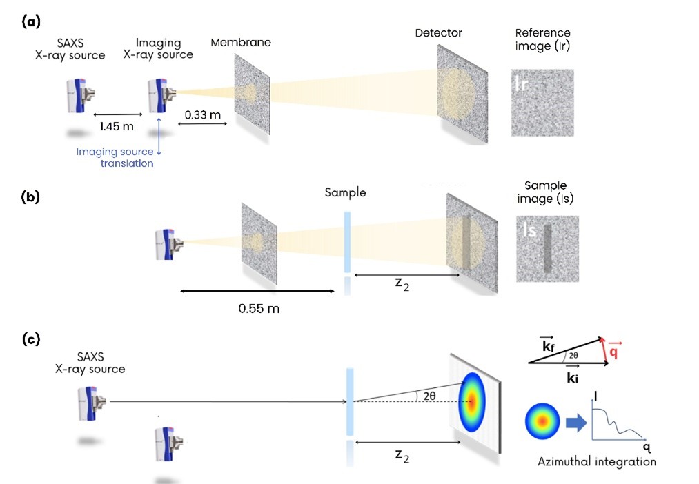
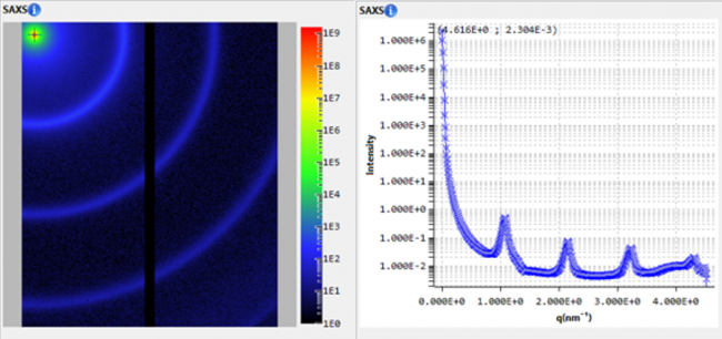
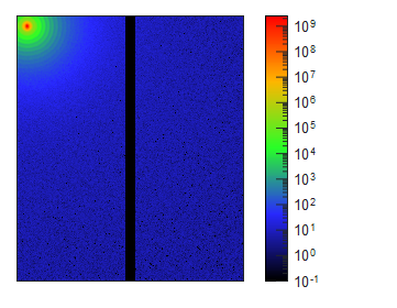
Figure 1. Dual-mode setup for combined dark-field imaging and SAXS on the Xeuss Pro. A membrane modulates the beam to generate phase and dark-field contrast (a,b), while SAXS measurements (c) record the scattered intensity I(q) from the same sample area. This configuration uniquely allows direct correlation between spatially resolved DF images and SAXS scattering curves.
While it is now widely accepted that the DF signal arises from X-ray scattering events caused by sub-resolution structures, the exact nature of these events and the angular sensitivity of the signal remain active topics of research. The simplest models attribute the origin of the dark-field signal solely to multiple refraction events within the sample [2, 3]. In contrast, more complex models attempt to directly relate the events measured in dark-field to small angle X-ray scattering (SAXS) intensity [4, 5].
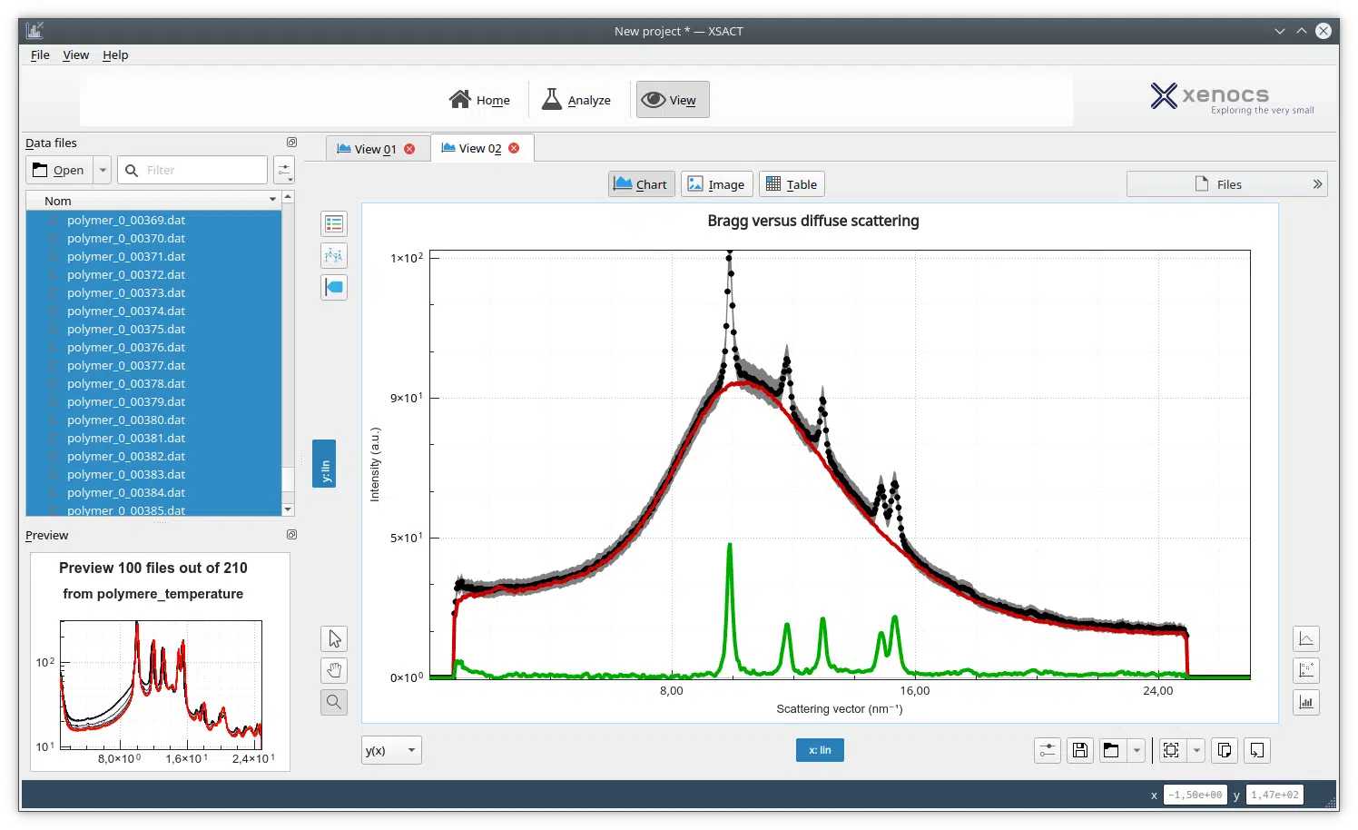
In this application note, you will discover how the Xeuss Pro combines dark-field imaging and small-angle X-ray scattering (SAXS) to provide complementary insights into nanoscale structure. This unique capability allows both techniques to be performed on the same sample without repositioning, offering correlated information on scattering intensity and spatial distribution.
Here, we use this combined approach to investigate the physical origins of the dark-field signal, specifically, which scattering events and angular ranges contribute to image contrast. By comparing dark-field images and SAXS curves acquired on identical materials, we show that dark-field contrast arises from both multiple refraction and small-angle scattering, and that its sensitivity primarily covers the low-q (USAXS) regime.
The results shown here are drawn from a broader investigation, with the full methodology and analysis available in [6].