Applied Surface Science, 2018, vol 436pp. 141-151
DOI:10.1016/j.apsusc.2017.11.218
Abstract
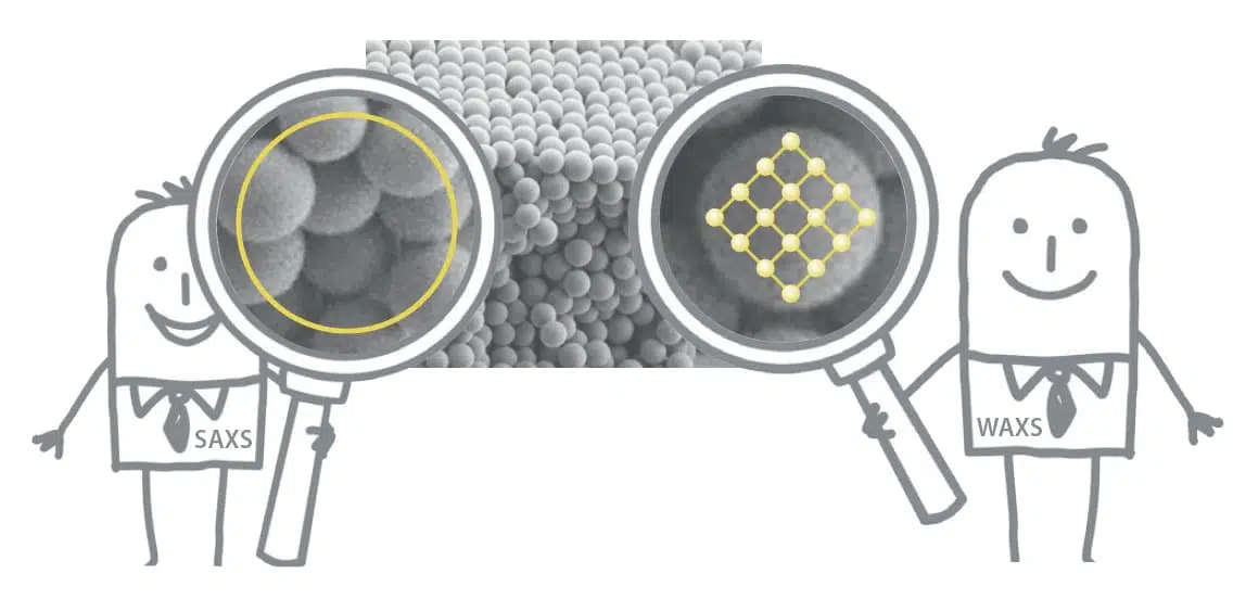
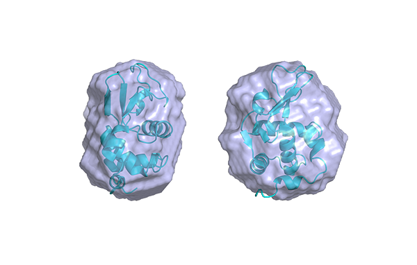
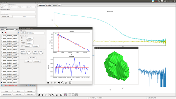
Hydroxyapatite is one of the most important biomaterials whose application mainly extends to implants and drug delivery. This work will discuss the changes in the pore size distribution of hydroxyapatite when there are latex beads present during the synthesis. These changes were monitored using different techniques: small angle X-ray scattering, X-ray diffraction, thermal gravimetrical analysis, N2 adsorption, scanning and transmission electron microscopy. Latex beads and hydroxyapatite form a single nanocomposite with well-distinguished inorganic and organic phases. Latex bead removal in the temperature range of 300–600 °C did not modify the original crystalline structure of hydroxyapatite. However, the latex beads favored an increase in the adsorption capacity of mesopores at temperatures higher than their glassy transition (Tg). The main result of this research work consists on the increase of surface area and pore size distribution obtained after the removal of latex beads template. Latex beads have been used in a different approach changing the porosity of hydroxyapatite scaffolds not only introducing new routes for cell integration but also broadening the pore size distribution which can result in a more high efficiency for drug release in living cells.