Calcified Tissue International, 2016, vol 99, 1, pp. 76-87
DOI:10.1007/s00223-016-0120-z
Abstract
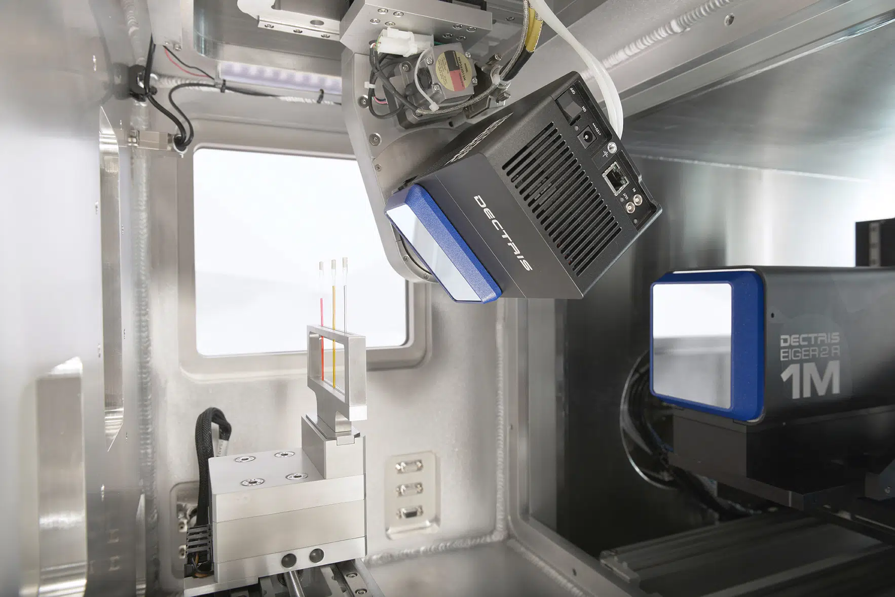
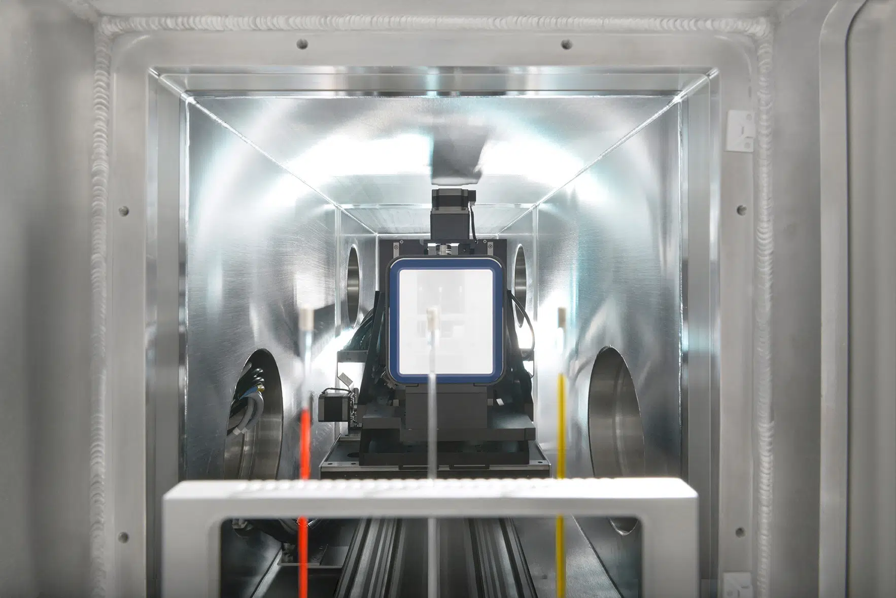
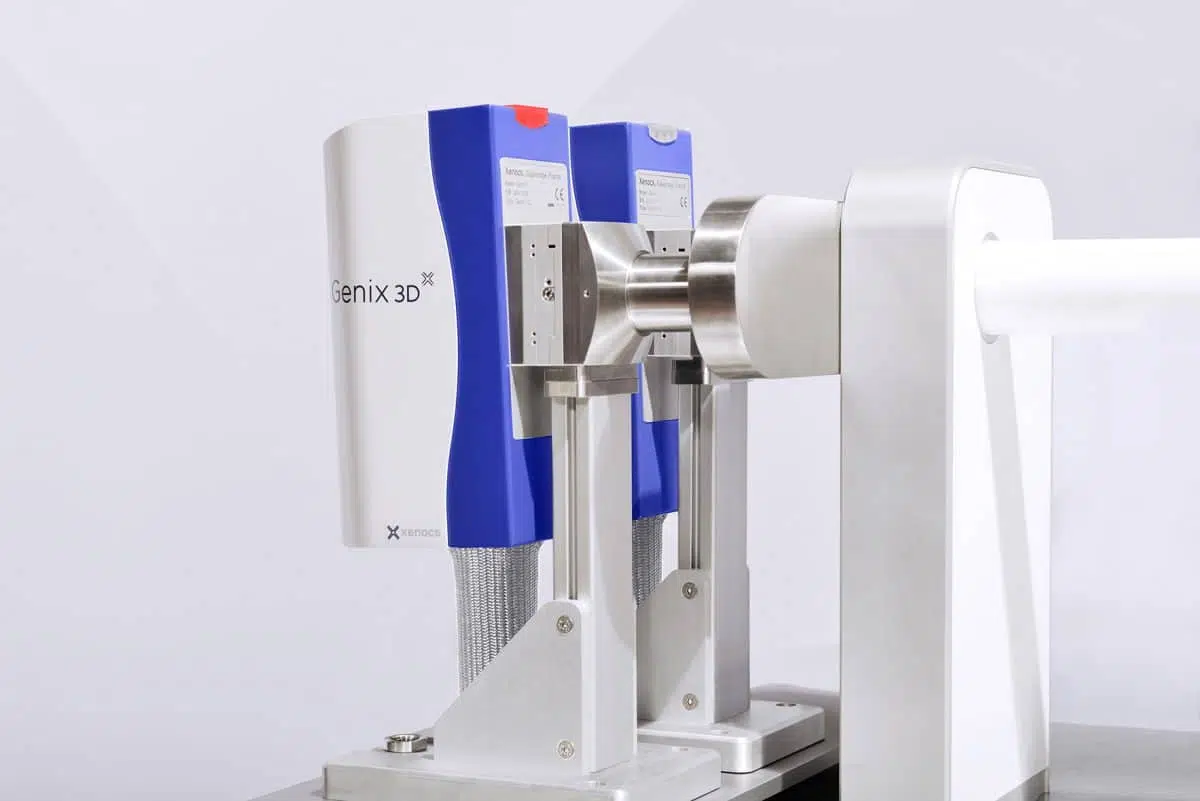
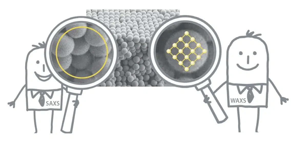
Despite the vast amount of studies focusing on bone nanostructure that have been performed for several decades, doubts regarding the detailed structure of the constituting hydroxyapatite crystal still exist. Different experimental techniques report somewhat different sizes and locations, possibly due to different requirements for the sample preparation. In this study, small- and wide-angle X-ray scattering is used to investigate the nanostructure of femur samples from young adult ovine, bovine, porcine, and murine cortical bone, including three different orthogonal directions relative to the long axis of the bone. The radially averaged scattering from all samples reveals a remarkable similarity in the entire q range, which indicates that the nanostructure is essentially the same in all species. Small differences in the data from different directions confirm that the crystals are elongated in the [001] direction and that this direction is parallel to the long axis of the bone. A model consisting of thin plates is successfully employed to describe the scattering and extract the plate thicknesses, which are found to be in the range of 20–40 Å for most samples but 40–60 Å for the cow samples. It is demonstrated that the mineral plates have a large degree of polydispersity in plate thickness. Additionally, and equally importantly, the scattering data and the model are critically evaluated in terms of model uncertainties and overall information content.