Chemistry of Materials, 2016, vol 28, 2, pp. 501-510
DOI:10.1021/acs.chemmater.5b03295
Abstract
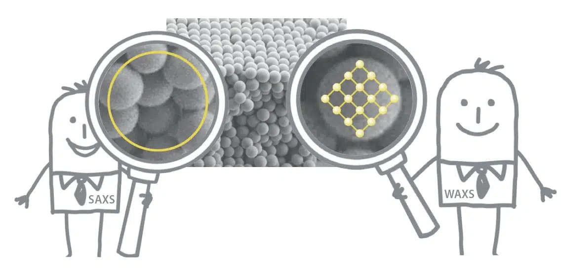
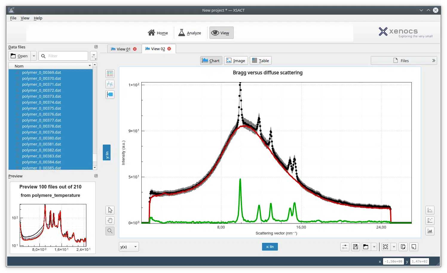
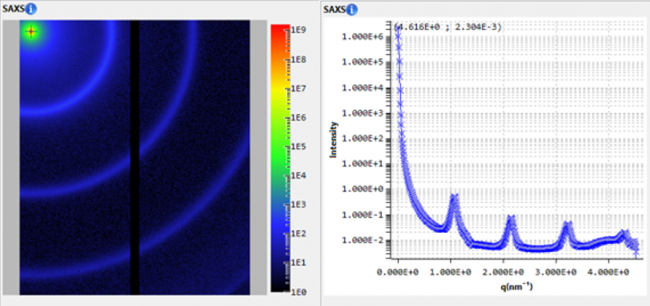
We describe the room-temperature synthesis and characterization of needle-like lanthanide phosphate (LnPO4) nanocrystals in water, based on in situ precipitation of LnPO4 using functional aqueous microgels as a soft nanoreactor. The poly(NIPAm/VCL/MAA) microgels were prepared by the copolymerization of N-isopropylacrylamide, N-vinylcaprolactam, and methacrylic acid. Our goal was to prepare Ln-encoded microgels suitable for bead-based biological assays employing mass cytometry. The low solubility of nanocrystalline LnPO4 avoids the problem of Ln ion leakage from the microgels. The main challenge was to find appropriate conditions to confine the LnPO4 precipitate to the interior of the microgels. Various sources of phosphate ions led to precipitation of LnPO4 on the exterior of the microgels. One approach worked. It involved three steps: neutralization of the MAA groups in the microgels with NaOH, ion exchange with a lanthanide salt, followed by treatment with a large excess of PBS buffer at pH 7.4. In this way, we obtained microgels containing ca. 107 Ln atoms per microgel (Ln = La, Nd, Eu, Tb, Ho, Tm). The microgels were shown to be uniform in size by dynamic light scattering and transmission electron microscopy. The LnPO4-containing microgels showed much higher stability against leakage of metals to acidic buffers compared to the LnF3-containing microgels reported previously [Lin, W.; Langmuir 2011, 27, 7265]. Small-angle X-ray scattering measurements and selected area electron diffraction data showed striking differences in the internal structure of hybrid microgels containing TbPO4 from those containing TbF3. The TbF3-containing microgels had a core?shell structure with an amorphous TbF3 core, whereas the TbPO4 microgels contained needle-like nanocrystals (width ca. 2 nm, lengths on the order of 80 nm) distributed throughout the structure. These microgels are very promising materials for biological assays based on mass cytometry.