
A novel spiral-patterned membrane enables superior resolution and contrast in single-mask X-ray phase contrast and dark-field imaging

X-ray imaging techniques based on phase contrast and dark-field signals offer rich structural insights beyond conventional absorption imaging. These methods are particularly valuable in visualizing features such as soft matter interfaces, microstructural orientations, and low-density variations in materials. One promising class of techniques—single-mask or membrane-based imaging—simplifies implementation by shifting experimental complexity into digital processing. This makes them well-suited for integration into laboratory X-ray systems such as the Dark Field and Phase Contrast Imaging (DF-PCI) option developed by Xenocs.

In this method, a beam modulator (mask or membrane) is placed in the X-ray beam to produce a structured pattern on the detector. A reference image is first recorded with the membrane alone, followed by a second image after insertion of the sample (as exemplified in Figure 1). The sample locally distorts the reference pattern by attenuating, deflecting and scattering the X-rays. Several pairs of reference and sample images are acquired while shifting the membrane position. By comparing how the sample modifies the reference pattern, it becomes possible to reconstruct attenuation, phase contrast and dark-field images. For the latter, the anisotropy in the scattering signal can also be derived from the reference/sample comparison.

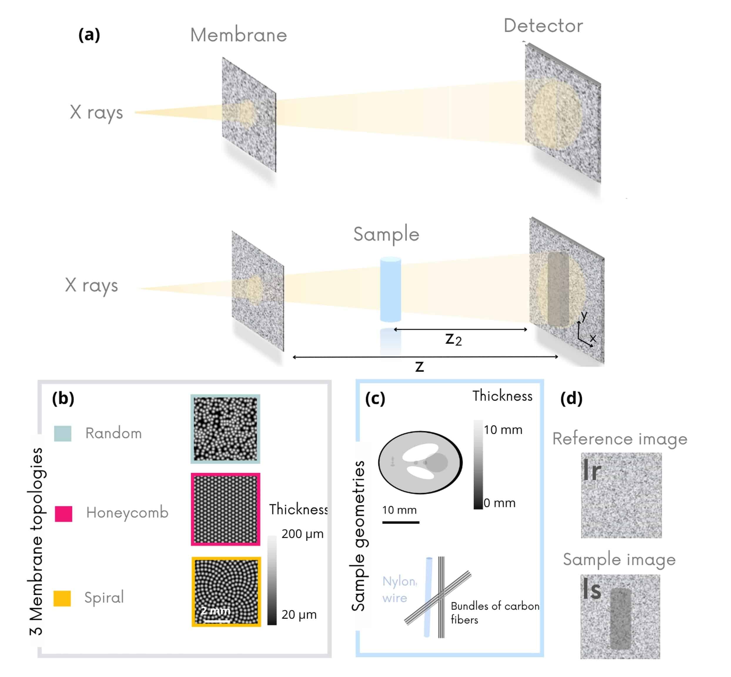
Figure 1. (a, d) Modulation Based imaging (MoBI) method consists in inserting a single mask into the X-ray beam to locally modulate the intensity on the detector. Several pairs of images are acquired of the mask alone (Ir) and of the mask with the sample (Is) by shifting the mask position between each acquisition pair. b) Three different modulation topologies (c) and two types of samples have been compared in this application note.

X-ray imaging techniques based on phase contrast and dark-field signals offer rich structural insights beyond conventional absorption imaging. These methods are particularly valuable in visualizing features such as soft matter interfaces, microstructural orientations, and low-density variations in materials. One promising class of techniques—single-mask or membrane-based imaging—simplifies implementation by shifting experimental complexity into digital processing. This makes them well-suited for integration into laboratory X-ray systems such as the Dark Field and Phase Contrast Imaging (DF-PCI) option developed by Xenocs.

The geometry of the membrane used to modulate the beam plays a critical role in determining the image quality. Each of these has trade-offs in terms of signal uniformity, modulation directionality, and susceptibility to artifacts. In this context, Xenocs has developed and patented a new membrane design based on a spiral geometry, inspired by Vogel patterns, which corresponds to the optimal balance between isotropy and modulation density.

In this application note, we introduce the working principle of the single-mask method and present results from simulations and experimental measurements comparing three membrane topologies: random, hexagonal, and spiral. Regular grid patterns were excluded from the analysis due to their tendency to introduce strong artifacts caused by their directional structure. We describe how these membranes impact image quality using key metrics such as signal-to-noise ratio, resolution, and structural similarity, and explain why the spiral membrane design provides superior imaging performance in both phase and dark-field contrast.

The results shown here are drawn from a broader investigation of membrane geometries, with the full methodology and analysis available in [8].

Author: C. Magnin
Measurements and Data Analysis: C. Magnin
Scientific reviewers: B. Faure, A. Tutueanu
References
[1] S. Berujon, H. Wang, and K. Sawhney. X-ray multimodal imaging using a random-phase object. Physical Review A 86, 063813 (2012). DOI: 10.1103/PhysRevA.86.063813
[2] V. Di Trapani, S. Savatović, F. De Marco, G. Lautizi, M. Margini, and P. Thibault. Speckle-based imaging (SBI) applications with spectral photon counting detectors at the newly established OPTIMATO (OPTimal IMAging and TOmography) laboratory. Journal of Instrumentation 19, C01018 (2024). DOI: 10.1088/1748-0221/19/01/C01018
[3] M.-C. Zdora, P. Thibault, T. Zhou, F. J. Koch, J. Romell, S. Sala, A. Last, C. Rau, and I. Zanette. X-ray phase-contrast imaging and metrology through unified modulated pattern analysis. Physical Review Letters 118, 203903 (2017). DOI: 10.1103/PhysRevLett.118.203903
[4] H. H. Wen, E. E. Bennett, R. Kopace, A.F. Stein, and V. Pai. Single-shot x-ray differential phase-contrast and diffraction imaging using two-dimensional transmission gratings. Optics Letters 35, 1932–1934 (2010). DOI: 10.1364/OL.35.001932
[5] Y. Y. How, D. M. Paganin, and K.S. Morgan. On the quantification of sample microstructure using single-exposure x-ray dark-field imaging via a single-grid setup. Scientific Reports 13, 11001 (2023). DOI: 10.1038/s41598-023-38199-z
[6] L. Guitard, A. Stolidi, G. Giakoumakis, R. Sousa Martins, J. Primot, and A. Jarnac. Robust quantitative X-ray phase diagnostic for carbon composite characterisation in the context of lightning induced risk. Scientific Reports 14, 21803 (2024). DOI: 10.1038/s41598-024-72087-7
[7] M. K. Croughan, Y. Y. How, A. Pennings, and K. S. Morgan. Directional dark-field retrieval with single-grid x-ray imaging. Optics Express 31, 11578–11597 (2023). DOI: 10.1364/OE.480031
[8] C. Magnin, L. Quenot, D. M. Cenda, B. Lantz, B. Faure, and E. Brun. On the optimisation of the geometric pattern for structured illumination based X-ray phase contrast and dark field imaging: A simulation study and its experimental validation. (2024). DOI: 10.48550/arXiv.2504.10665