WEBINAR
New contrasts in full-field X-ray imaging and
their complementarity with SAXS
Tuesday, October 21, 2025
15:00–15:45 CEST (UTC+2) | 9:00–9:45 AM EDT
Standard X-ray radiography often struggles to differentiate materials with similar absorption properties, limiting the level of insight it can provide. Phase-contrast (PC) imaging and dark-field (DF) imaging are innovative new X-ray imaging modalities that overcome these limitations and open new perspectives for sample characterization.
Phase-contrast imaging detects subtle refraction of X-rays within a sample, sharply enhancing interfaces and boundaries that are almost invisible in attenuation images.
Dark-field imaging is sensitive to ultra-small-angle scattering events, providing full-field maps of nanoscale heterogeneities and orientation effects.
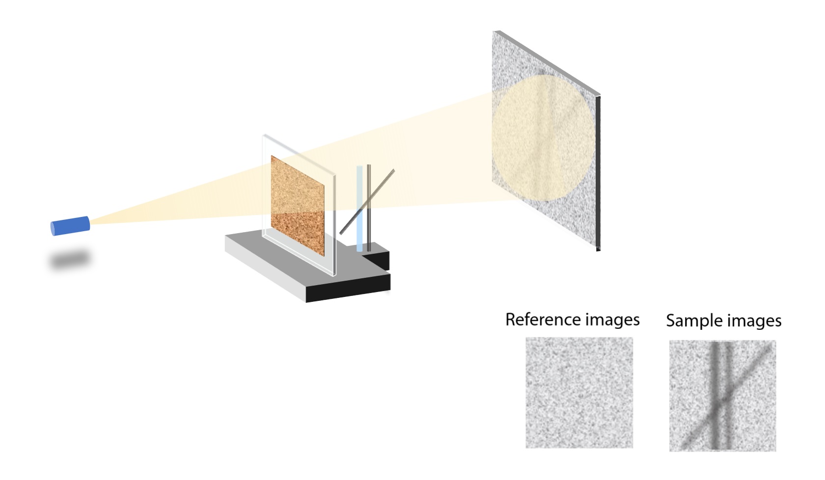
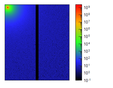
Figure 1: Schematic of a speckle-based X-ray imaging setup showing reference and sample images.
The combination of these two modalities enables researchers to reveal hidden structural features, distinguish between otherwise indistinguishable materials, and connect macroscopic visualization with nanometric detail.
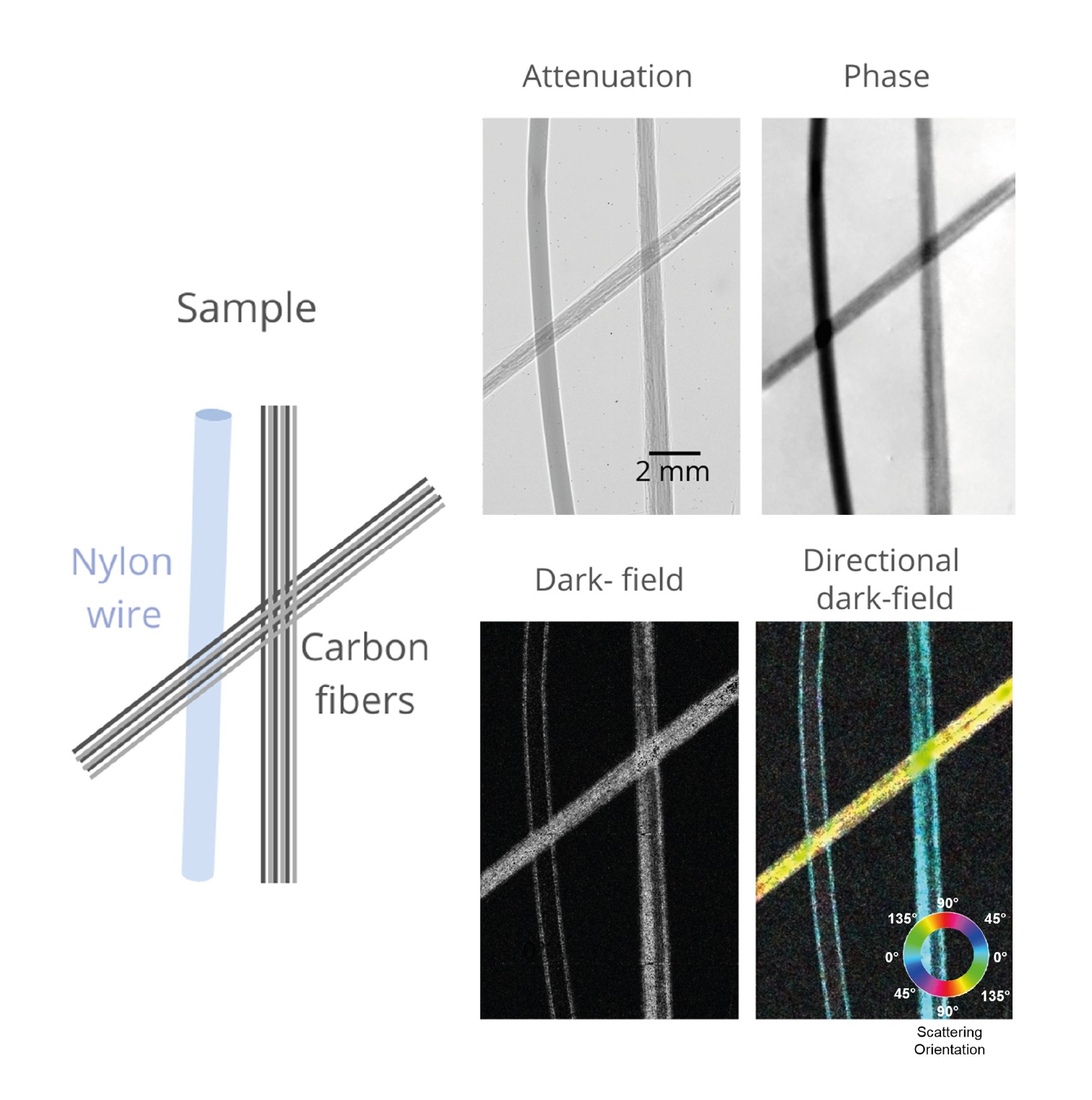
Figure 2: Attenuation (a), phase (b), dark-field (c), and directional dark-field (d) images of a nylon wire and carbon fiber bundles. While attenuation imaging does not distinguish the materials, phase-contrast and dark-field imaging highlight differences in composition, nanostructure, and orientation.
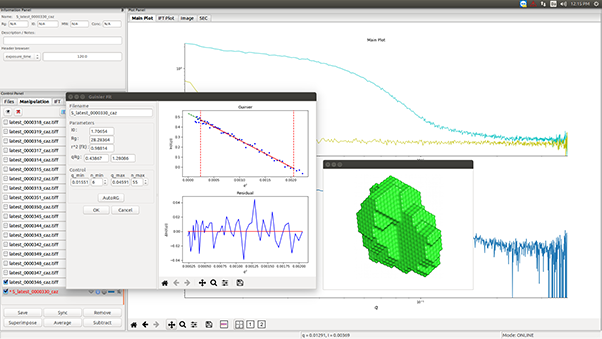
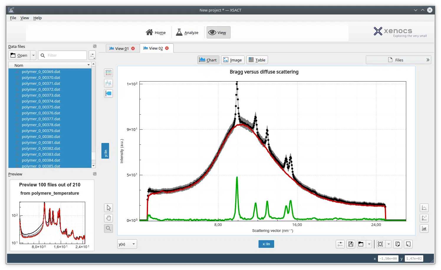
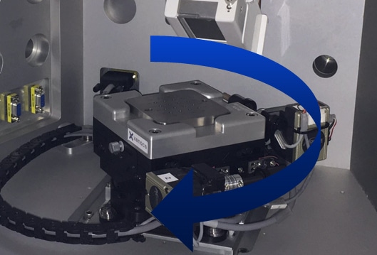
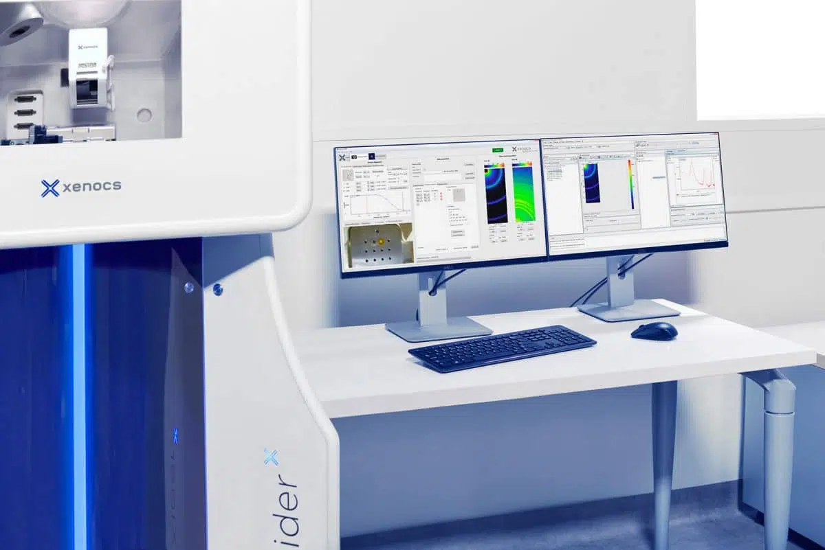
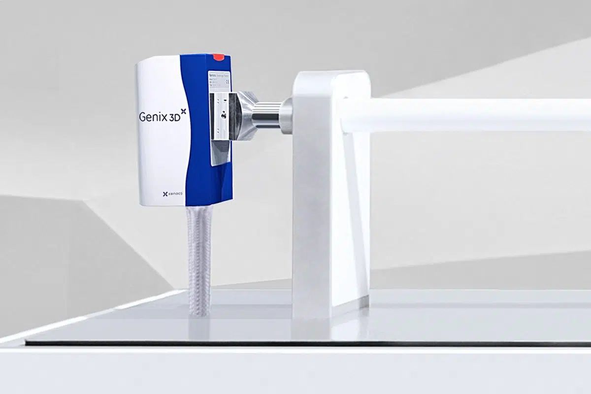
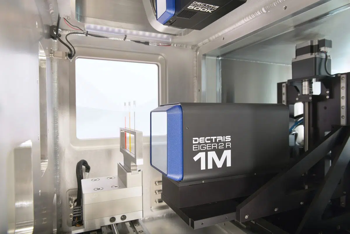
The DF-PCI option developed by Xenocs on the Xeuss instruments extends these imaging capabilities even further by uniquely integrating them with SAXS. This makes it possible to correlate full-field contrasts with quantitative nanostructural information, providing a comprehensive understanding of materials across length scales.
Register now to learn:
- What phase-contrast and dark-field imaging reveal beyond conventional attenuation.
- The advantages of these modalities for distinguishing and analyzing complex materials.
- How dark-field imaging can guide SAXS measurements and enhance their interpretation.
- Case studies that demonstrate the added value of combining SAXS with advanced imaging.
Presented by:

Clara Magnin holds a PhD in X-ray imaging from the Strobe laboratory (INSERM), conducted in collaboration with Xenocs. After an academic background in fundamental physics and a Master’s specialization in medical physics, her doctoral research focused on the development of new X-ray imaging methods. Her PhD work aimed to transfer innovative X-ray imaging technologies developed at synchrotron facilities to laboratory-based sources. She developed phase-contrast and dark-field imaging methods for low-coherence systems. One of the main research directions of her thesis involved understanding the scattering signal measured in dark-field imaging and exploring the connection between this modality and small-angle X-ray scattering (SAXS).
Organizer

Ana Tutueanu, PhD
Product Marketing and Scientific Communication Specialist
Email: [email protected]