RSC Advances, 2016, vol 6, 67, pp. 62119-62127
DOI:10.1039/C6RA09616F
Abstract
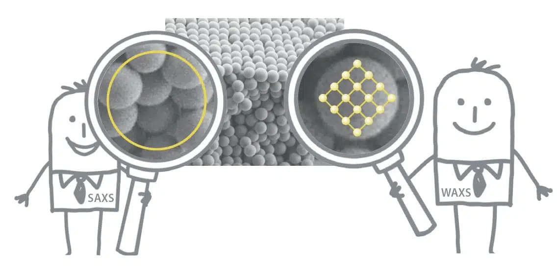
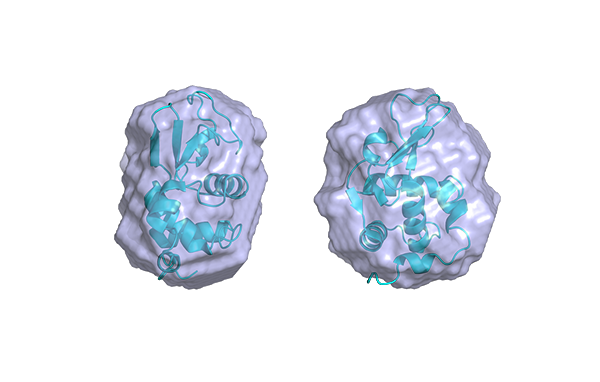
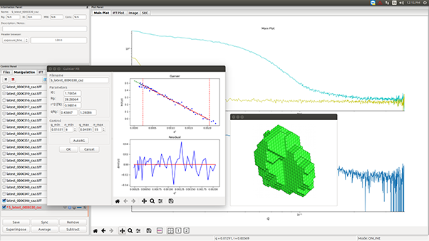
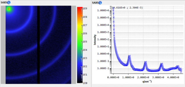
To evaluate the potential of monoolein-based cubic bicontinuous liquid crystalline nanoparticles (cubosomes) as diagnostic tools in diabetes and pancreatic β-cell transplantation, fluorescent cubosomes were formulated, entrapping within the lipid bilayer a newly synthesized, potent, hydrophobic dye. Cryo-TEM, SAXS, and DLS were performed to assess the morphological and structural aspects of this formulation. Rat pancreatic β-cells (INS-1E) readily took up the cubosome nanoparticles, as revealed by in vitro internalization tests, and flow-cytometric analyses confirmed the persistence of the fluorescence for up to 24 hours. The cytotoxicity of cubosomes at various concentrations, evaluated with a real-time analysis of proliferation, revealed that the proliferation curves relative to the less concentrated formulation (5 μM in terms of monoolein content) were comparable to the untreated cultures. INS-1E cell cycle and apoptosis supported such results. In conclusion, this formulation, with dye content in the nanomolar range, seems to be useful for β-cell imaging.